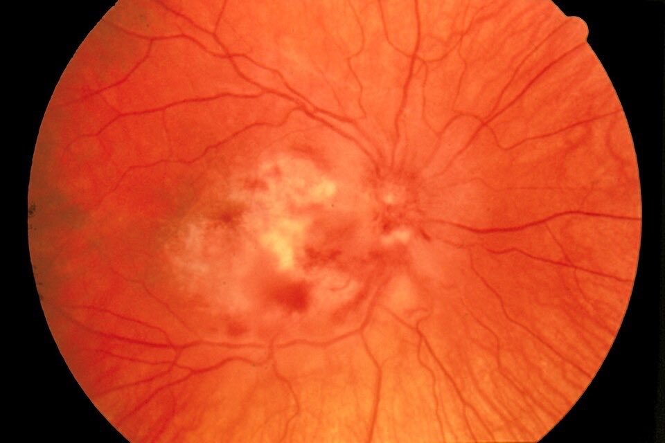
Q. An HIV-positive patient presents with painless, progressive loss of vision. Fundus examination findings are seen in the image shared above. What is the most likely diagnosis?
A) Diabetic retinopathy.
B) CMV retinitis.
C) Hypertensive retinopathy.
D) Retinal detachment.
✓ The Correct Answer is: B) CMV Retinitis.
Cytomegalovirus (CMV) retinitis is an important opportunistic infection seen in patients with advanced HIV infection, particularly those with severe immunosuppression.
It typically causes painless, progressive visual impairment, although floaters and visual field defects may also occur.
The fundus appearance shown in the image is characteristic of CMV retinitis, with areas of necrotizing retinitis and retinal hemorrhage. The classic description is “pizza pie” or “cottage cheese and ketchup” appearance.
∆ Clinical Features
CMV retinitis may present with:
1. Painless progressive loss of vision.
2. Floaters or blurred vision.
3. Scotomas or visual field defects.
4. Retinal whitening due to necrosis.
5. Associated retinal hemorrhages.
The disease commonly occurs in patients with advanced HIV, especially when the CD4 count is very low (classically <50 cells/µL).
∆ Diagnosis
Diagnosis is usually made clinically by dilated fundus examination demonstrating characteristic necrotizing retinitis. In atypical cases, ocular fluid testing for CMV DNA by PCR can help confirm the diagnosis.
∆ Management in Clinical Practice
Treatment involves systemic anti-CMV therapy, commonly with valganciclovir or intravenous ganciclovir, depending on the clinical situation.
Sight-threatening lesions may require intravitreal antiviral therapy in addition to systemic treatment.
An equally important part of management is effective antiretroviral therapy (ART) to restore immune function and reduce the risk of recurrence.
∆ Other options:
Diabetic retinopathy: Associated with diabetes and typically produces microaneurysms, hemorrhages, exudates, and/or neovascularization rather than necrotizing retinitis.
Hypertensive retinopathy: Characterized by arteriolar narrowing, AV crossing changes, hemorrhages, cotton-wool spots, and sometimes papilledema in severe disease.
Retinal detachment: Usually presents with flashes, floaters, and a curtain-like visual-field defect rather than the characteristic necrotizing retinal lesions seen in this image.
∆ High-Yield Exam Pearls:
HIV + painless progressive visual loss → Think CMV retinitis.
Classic fundus: “Pizza pie”/“cottage cheese and ketchup” appearance.
Typical setting: Advanced HIV with severe immunosuppression.
Classic CD4 association: <50 cells/µL.
Diagnosis: Characteristic fundus examination; ocular PCR can assist in atypical cases.
Treatment: Systemic anti-CMV therapy + optimization of ART.
✓ One-Liner to Remember:
“Advanced HIV + painless visual loss + hemorrhagic necrotizing retinitis = CMV retinitis.”
✓ Take-Home Message:
CMV retinitis should be considered whenever a patient with advanced HIV develops painless, progressive visual symptoms.
Recognizing the characteristic necrotizing, hemorrhagic retinal lesions allows prompt diagnosis and treatment, which is crucial because untreated CMV retinitis can result in permanent visual loss.
This classic association is highly relevant for MBBS, NEET PG, INICET, FMGE, USMLE, PLAB and other medical examinations, as well as clinical practice.